Researchers at the University of Michigan have developed a groundbreaking artificial intelligence system capable of interpreting brain MRI scans in mere seconds. The model, named Prima, achieved diagnostic accuracy as high as 97.5% across more than 50 different neurological conditions. By automatically identifying critical cases like strokes and brain hemorrhages, the tool is designed to help radiologists prioritize life-threatening emergencies and reduce dangerous bottlenecks in patient care.
This new technology, detailed in a study published in Nature Biomedical Engineering, represents a significant shift in medical imaging. Unlike previous AI models trained for narrow tasks—such as detecting a single type of tumor—Prima functions as a generalist. It can recognize a broad spectrum of disorders, from aneurysms to hydrocephalus, and even assess the urgency of a patient’s condition immediately after imaging is complete.
Addressing the Radiologist Shortage
The development of Prima comes at a time when the demand for MRI scans is outpacing the availability of specialists trained to read them. This imbalance often leads to delays in diagnosis, which can be detrimental for patients suffering from acute neurological issues. In rural hospitals or overburdened health systems, it can sometimes take days for a scan to be interpreted by a neuroradiologist.
“As the global demand for MRI rises and places significant strain on our physicians and health systems, our AI model has the potential to reduce burden by improving diagnosis and treatment with fast, accurate information,” said Todd Hollon, M.D., the study’s senior author and a neurosurgeon at University of Michigan Health.
By acting as an intelligent triage system, Prima aims to close this gap. When the model detects an acute issue, it can instantly alert the appropriate subspecialist, such as a stroke neurologist or neurosurgeon, ensuring that time-sensitive conditions are treated without unnecessary administrative delays.
How Prima Works
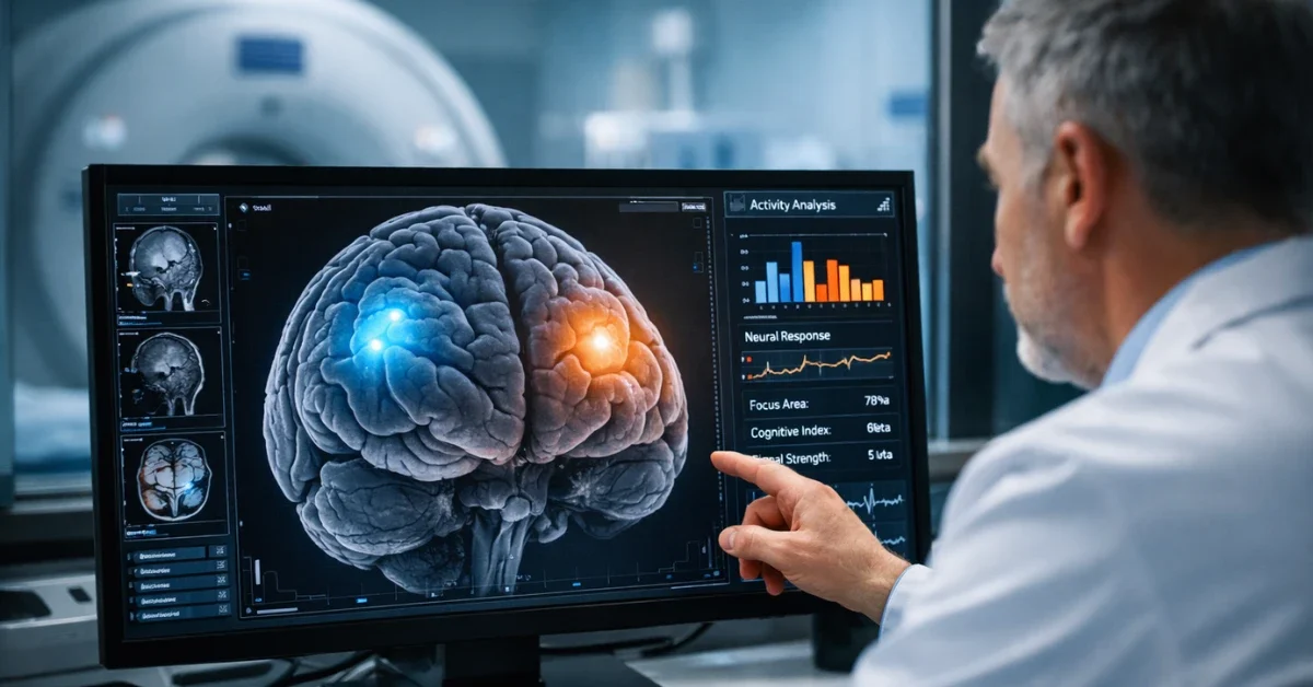
Prima is classified as a vision-language model (VLM), a type of AI architecture similar to the technology behind chatbots like ChatGPT, but adapted for medical data. It processes images, video sequences, and text simultaneously to generate a comprehensive understanding of a patient’s health.
To build the system, researchers trained Prima on a massive dataset comprising more than 200,000 MRI studies and 5.6 million individual imaging sequences—essentially every MRI collected at University of Michigan Health since radiology records were digitized. Crucially, the model was also trained on patients’ clinical histories and the specific reasons physicians ordered the scans. This allows Prima to interpret images within the context of a patient’s medical background, mirroring the workflow of a human radiologist.
“Prima works like a radiologist by integrating information regarding the patient’s medical history and imaging data to produce a comprehensive understanding of their health,” said co-first author Samir Harake, a data scientist in the Machine Learning in Neurosurgery Lab.
Performance and Future Potential
In testing, the research team evaluated Prima using more than 30,000 real-world MRI studies over a one-year period. The results showed that Prima outperformed existing advanced AI models in both diagnostic accuracy and its ability to prioritize urgent cases. Its success in identifying diverse conditions suggests that training on large-scale, real-world data is more effective than using smaller, curated datasets.
While the results are promising, the researchers emphasize that Prima is currently in an early evaluation phase and is intended to serve as a “co-pilot” for physicians rather than a replacement. The goal is to streamline clinical workflows and improve access to expert-level radiology services, regardless of where a patient is treated.
“Whether you are receiving a scan at a larger health system that is facing increasing volume or a rural hospital with limited resources, innovative technologies are needed to improve access to radiology services,” said Vikas Gulani, M.D., Ph.D., chair of the Department of Radiology at U-M Health.
Future research will focus on integrating even more detailed patient data from electronic medical records to further refine the model’s accuracy. The team also believes the underlying technology could eventually be adapted for other types of medical imaging, such as chest X-rays, mammograms, and ultrasounds.
